What Is Pelvic Venous Disorder (PeVD)?
Pelvic Venous Disorder (PeVD) is a vascular condition involving abnormal blood flow within the veins of the pelvis. It can present with a range of symptoms—most commonly a persistent, dull pelvic ache—that may fluctuate with posture, menstrual cycle, or daily activity. Because these symptoms overlap with other conditions, PeVD is often not immediately recognised.
Why Standard Imaging May Not Detect It
A key feature of PeVD is that symptoms are related to how blood flows through the pelvic veins rather than to obvious structural changes. As a result, routine imaging such as standard ultrasound or MRI, which primarily assess anatomy, may not always identify the underlying issue. Diagnosis typically requires a specialised duplex ultrasound that evaluates venous flow and valve function in real time.
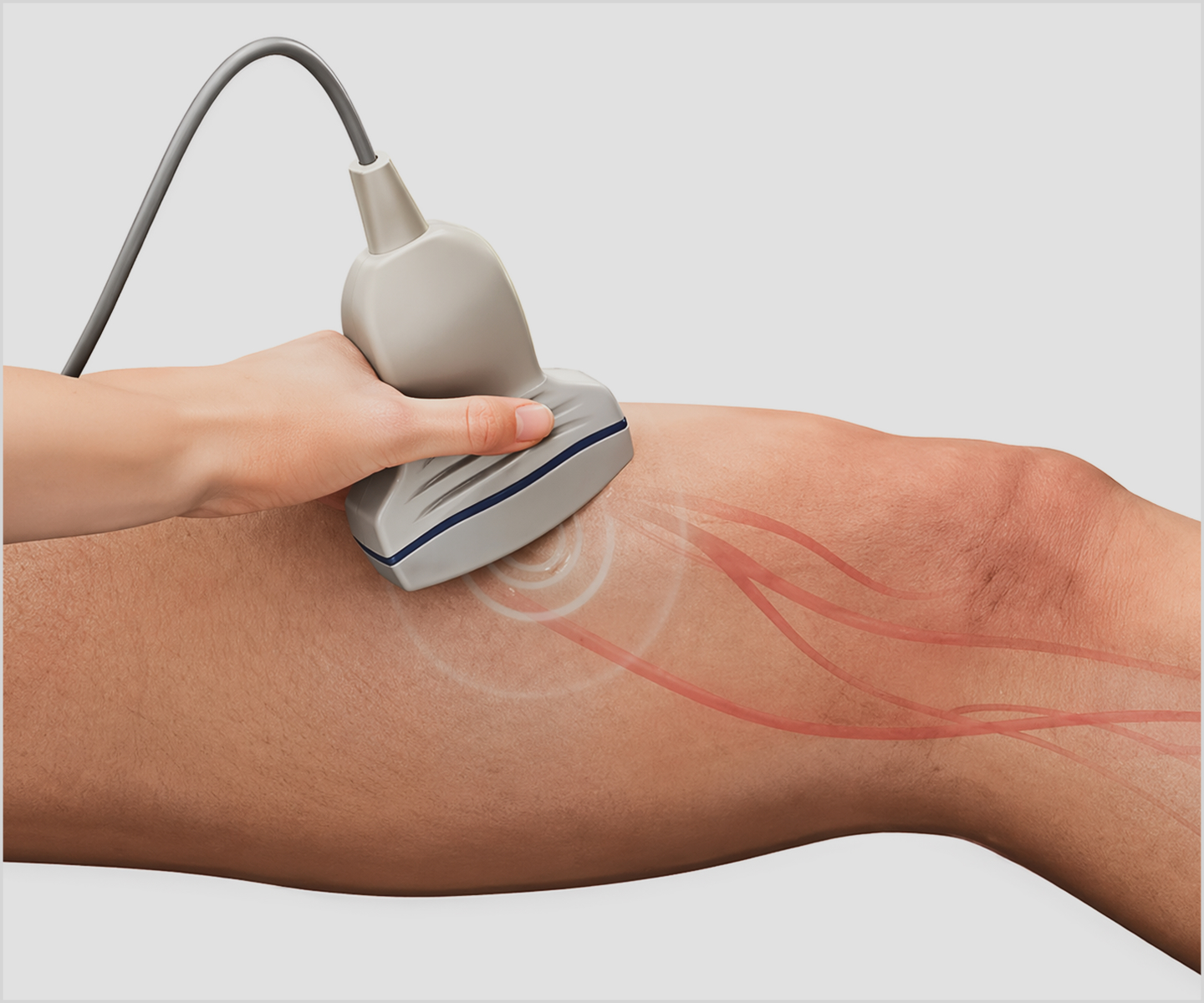
Specialised duplex ultrasound
A key reason PeVD can be missed is that most routine imaging is designed to capture static anatomy, not dynamic blood flow. Standard ultrasound and MRI are typically performed with the patient lying flat, a position that can reduce venous pooling and temporarily normalise vein appearance. In contrast, PeVD symptoms are often driven by venous reflux or impaired drainage that becomes more evident when upright or after prolonged activity—conditions not replicated during routine scans.
In addition, conventional imaging may not consistently assess vein diameter changes over time, direction of blood flow, or valve competence, all of which are central to identifying venous dysfunction. Pelvic veins can also be variable and complex in their anatomy, making subtle abnormalities harder to interpret without a targeted protocol.
Specialised duplex ultrasound addresses these gaps by combining real-time imaging with Doppler flow assessment. This allows clinicians to evaluate how blood moves through the pelvic veins under different conditions, helping to detect reflux patterns or flow disturbances that standard imaging may not capture.
How Symptoms Can Vary
Women may experience PeVD differently. These variations can make the condition difficult to identify without a focused assessment.
- Some describe a feeling of heaviness or pressure in the lower abdomen.
- Others notice discomfort after prolonged standing or at the end of the day.
- Symptoms can also be influenced by hormonal changes, including after pregnancy.
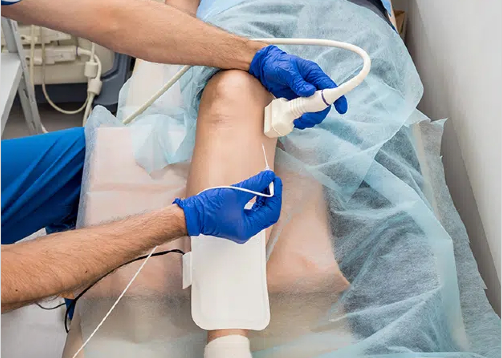
Diagnosis and Care in Singapore
In Singapore, access to specialised vascular imaging and multidisciplinary care can support a more structured approach to evaluation. This often includes a detailed clinical history, symptom mapping, and targeted imaging to better understand individual patterns. Management is tailored accordingly and may involve conservative measures, minimally invasive procedures, or supportive therapies, depending on clinical findings and patient preferences.
Considering the Next Step
If these symptoms feel familiar, you may wish to explore them further using a self-assessment tool or by arranging a clinical evaluation.
This can help clarify whether PeVD may be contributing to your experience and what next steps, if any, could be appropriate.