Pelvic Vein Embolisation in Singapore: A Minimally Invasive Treatment for Pelvic Venous Disorder

Performed at The Venus Clinic in Singapore by vascular specialist Dr Sriram Narayanan, pelvic vein embolisation is a minimally invasive, image-guided treatment for pelvic venous disorder (PeVD).

If you have been living with chronic pelvic heaviness, unexplained varicose veins around your thighs, or deep discomfort that worsens after standing, you may be dealing with pelvic venous insufficiency. Often described as “varicose veins in the pelvis,” this vascular condition may be treated in Singapore with a procedure called pelvic vein embolisation.

4a6dbe57364c4538806f1a8bca876dac721f8d25

How Pelvic Vein Embolisation Works

To understand how this procedure helps, it is useful to first understand what occurs at the vascular level.
Frame 1973

Your pelvic veins have a job.

They drain venous blood away from the uterus, ovaries, and surrounding pelvic structures back toward the heart. Inside these veins, small one-way valves prevent blood from flowing backwards. When these valves become incompetent (meaning they no longer close properly), blood begins to flow in the wrong direction. This is called retrograde flow, or reflux.

Frame 1974

The result is venous pooling.

Blood accumulates in the ovarian veins and their pelvic tributaries, causing them to dilate and become engorged. This creates sustained venous hypertension (raised blood pressure within the pelvic veins). Over time, this increased pressure leads to the chronic aching, heaviness, and fullness that characterise PeVD.

Frame 1974 (1)

Embolisation works by deliberately sealing These dysfunctional vessels.

A vascular surgeon navigates a catheter (a thin, flexible tube) into the incompetent veins and deploys embolic agents, such as metallic coils or sclerosant solutions, to close them. Once sealed, these veins can no longer carry refluxing blood. The body then naturally redirects venous drainage through healthy, competent pathways.

Frame 2147223875

The result is a reduction in pelvic venous pressure.

With less pooling and engorgement, the mechanical source of symptoms is addressed at its origin.
woman-relaxing-on-couch-with-hand-on-stomach-2026-01-16-17-46-02-utc (1) woman-relaxing-on-couch-with-hand-on-stomach-2026-01-16-17-46-02-utc (1)

Who Is a Suitable Candidate for Pelvic Vein Embolisation?

Not everyone with pelvic pain will require embolisation. Candidacy depends on a careful evaluation of your symptoms, their duration, and a confirmed vascular cause.

Symptom patterns that suggest a pelvic venous origin include:

  • Chronic pelvic pain lasting six months or longer that worsens after prolonged standing, walking, or physical activity
  • A deep, dragging heaviness in the lower abdomen or pelvis that eases when you lie down
  • Pain during or after sexual intercourse that lingers for hours
  • Visible varicose veins around the vulva, inner thighs, or buttocks that cannot be explained by standard lower-limb venous disease
  • A history of symptoms that began during or after pregnancy, or that have progressively worsened with successive pregnancies, noting that PeVD can also occur in women who have not been pregnant.

If three or more of these patterns sound familiar, a specialist vascular assessment can confirm whether pelvic venous disorder is the underlying cause.

Make An Enquiry
Frame 3540 Hero - Mobile (2)

The importance of exclusion

Many gynaecological and urological conditions share overlapping symptoms with PeVD. Endometriosis, uterine fibroids, ovarian cysts, interstitial cystitis, and musculoskeletal pelvic floor dysfunction can all produce chronic pelvic discomfort. Before embolisation is considered, these conditions must be assessed and either excluded or identified as co-existing factors.

Dr Sriram Narayanan, MBBS, MS (Surgery), FRCS (Glasgow), FRCS (Gen Surg), Dip Lap Surgery (France) — a Senior Consultant Vascular and Endovascular Surgeon practising at Gleneagles Hospital and Mount Elizabeth Novena Hospital — leads The Venus Clinic team with a comprehensive approach that addresses both the medical and psychosocial dimensions of pelvic venous disorder. His evaluation protocol integrates pelvic floor assessments alongside vascular imaging to ensure the full clinical picture is understood before any procedural decision is made. Dr Narayanan brings over two decades of vascular and endovascular surgical experience across the United Kingdom and Singapore to every patient assessment.

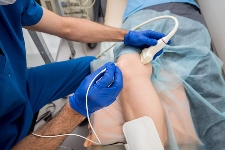
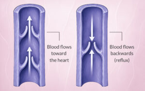
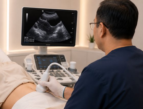
Definitive candidacy requires specialist imaging. Specialist imaging is required to confirm the presence and pattern of pelvic venous reflux before proceeding. Duplex ultrasound is commonly used as an initial screening tool, while cross-sectional venous imaging (CT or MR venography) and diagnostic venography provide more definitive anatomical detail, with catheter-directed venography considered the gold standard for diagnosis and often performed immediately prior to treatment.

Experiencing chronic pelvic pain that worsens with standing or prolonged activity?

A specialist vascular assessment can deliver diagnostic clarity on whether pelvic venous insufficiency is the confirmed cause. No GP referral is required, and every enquiry is reviewed by our team directly, not a call centre. Contact us today and speak with a vascular specialist within 48 hours.

Preparing for Your Pelvic Vein Embolisation Procedure

Preparation centres on diagnostic rigour. The goal is to confirm that your symptoms have a treatable vascular cause and to map the exact anatomy of your pelvic venous reflux before any intervention begins.

The diagnostic pathway typically includes:

  • Duplex ultrasonography: A non-invasive ultrasound study that detects dilated pelvic veins and reversed blood flow. Transabdominal or transvaginal approaches may be used. Dr Sriram Narayanan applies this as a foundational tool to identify venous reflux patterns and guide treatment planning.
  • Cross-sectional venous imaging: Dedicated venous-phase CT or MR venography provides detailed anatomical maps of the ovarian veins, internal iliac tributaries, and any variant anatomy that may influence the procedural approach.
  • Diagnostic venography: Catheter-directed venography is the gold standard for directly visualising and measuring reflux within the pelvic veins, providing definitive anatomical detail. It is typically performed as an immediate precursor to the treatment session itself, enabling diagnosis and intervention in a single procedure where appropriate.

What to expect before your procedure day

Your specialist will review your imaging findings, confirm the treatment plan, and discuss any medications that may need adjusting. You will typically be asked to fast for several hours beforehand. Bring a list of your current medications and any relevant imaging from prior assessments.

A note on expectations. Understanding that this procedure targets a specific vascular problem can help contextualise years of unexplained symptoms. A confirmed diagnosis provides a clinical explanation and a pathway forward.

Why thorough imaging matters. Many women spend years being told that their chronic pelvic pain is “just hormonal,” “stress-related,” or a normal part of their reproductive life. This means PeVD may be undiagnosed or misattributed to gynaecological conditions alone. A dedicated vascular assessment is essential to differentiate pelvic venous insufficiency from other conditions with similar symptom profiles.

What Happens During Pelvic Vein Embolisation?

The procedure is performed in a catheterisation laboratory (also called an interventional suite) equipped with fluoroscopic imaging. Here is what happens at each stage.

  • Venous access

    The specialist gains access through a tiny puncture, usually in the neck (internal jugular vein) or groin (femoral vein). Using the neck approach allows for a smoother, more direct path to the pelvic veins, often resulting in a faster procedure.

  • Catheter navigation

    Using real-time X-ray guidance (fluoroscopy), a thin catheter is advanced through the venous system toward the target pelvic veins, including the ovarian veins and any dilated internal iliac tributaries contributing to reflux.

  • Selective venography

    Once the catheter is positioned within the target vessel, contrast dye is injected to create a real-time map of the incompetent veins, their branches, and the direction of blood flow. This step confirms which vessels require treatment and identifies any collateral pathways.

  • Deployment of embolic agents

    The specialist deploys embolic materials to seal the dysfunctional veins. This may involve:

    • Metallic coils that mechanically block the vessel
    • Sclerosant agents that cause the vein wall to collapse and scar closed
    • A combination of both

    The choice of agent depends on vessel diameter, anatomy, and clinical judgement.

  • Haemodynamic confirmation

    After embolisation, additional venography confirms that reflux has been eliminated and that blood flow is appropriately redirected. The catheter is then removed, and gentle pressure is applied to the access site.

The entire procedure is conducted under continuous imaging guidance, allowing precise targeting and minimising the risk of non-target embolisation.

cropped-image-of-sick-woman-lying-on-hospital-bed-2026-01-06-00-40-00-utc (1) cropped-image-of-sick-woman-lying-on-hospital-bed-2026-01-06-00-40-00-utc (1)

Anaesthesia and Patient Comfort During the Procedure

Pelvic vein embolisation is typically performed under conscious sedation or local anaesthesia rather than general anaesthesia. You will be awake but relaxed throughout.

What you may feel. Some patients notice a sensation of warmth or mild pressure when contrast dye is injected. Brief cramping in the pelvis may occur as embolic agents are deployed, particularly if sclerosant is used. These sensations are expected and generally short-lived.

What to communicate. If you experience sharp pain, sudden chest discomfort, or difficulty breathing at any point, inform the procedural team immediately. Your vital signs are monitored continuously, and the sedation level can be adjusted to maintain your comfort.

The Recovery Journey After Pelvic Vein Embolisation


Time After Procedure What to Expect Activity Guidance
Day 0 (Procedure Day) Observed in recovery for a few hours post-procedure. Mild tenderness or bruising at the access site (neck or groin). Most patients discharged the same day; occasional overnight stay depending on complexity. Rest for the remainder of the day. Eat and drink normally after sedation wears off. A companion is advised for the journey home. Avoid driving on the day of the procedure.
Days 1–14 (Weeks 1–2) Pelvic aching or heaviness is a normal response as embolised veins begin to scar closed. Post-Embolisation Syndrome (temporary flu-like feeling, mild fever, or nausea) may occur and is usually self-limiting. Light daily activities can resume within a few days. Avoid heavy lifting, strenuous exercise, and prolonged standing. Report persistent fever, worsening pain, or any concerns to your care team promptly.
Weeks 2–6 Sealed veins continue to close fully; blood flow redistributes through healthy vessels. Some patients notice early symptom relief; others experience gradual improvement over weeks. Most patients can return to normal activity around one week post-procedure. More demanding activities should be resumed only on specialist advice.
Beyond 6 Weeks Continued symptom improvement expected; studies show sustained clinical benefit at 1, 2, and 3 years. Follow-up imaging confirms vein occlusion and checks for residual reflux. Full activity as cleared by your specialist. Attend your scheduled follow-up (typically around 6 weeks), which may include duplex ultrasound or other imaging to confirm vein occlusion and track symptom progress.
woman-suffering-from-stomach-pain-at-home-2026-03-17-21-42-40-utc (1) woman-suffering-from-stomach-pain-at-home-2026-03-17-21-42-40-utc (1)

When to Seek Urgent Medical Attention After the Procedure

While serious complications are uncommon, certain signs warrant immediate specialist contact:

  • Sudden severe pain in the pelvis, chest, or leg
  • Significant swelling, redness, or discharge at the access site
  • Fever above 38°C persisting beyond 24 to 48 hours, or any fever not consistent with expected post-embolisation syndrome
  • Difficulty breathing or chest tightness

These symptoms require prompt assessment to rule out events such as access-site infection, deep vein thrombosis, or coil migration. Contact your specialist team if something feels wrong.

For urgent concerns within 30 days of your procedure, contact The Venus Clinic on +65‎ 8666‎ 9639 or proceed to your nearest A&E if you experience severe pain, chest tightness, or difficulty breathing.’


Make An Enquiry

Concerned about your recovery or unsure whether your symptoms are improving as expected?

Structured post-procedural follow-up, including imaging review, provides objective confirmation of vascular occlusion and supports a clearly defined recovery pathway.

Other Treatment Options for Pelvic Congestion Syndrome


Treatment Anaesthesia Recovery Addresses Root Cause? Day Surgery?
Pelvic Vein Embolisation Local anaesthetic with conscious sedation Return to light activity within days; most resume normal activity ~1 week Yes — occludes incompetent veins directly Yes — same-day discharge in most cases
Hormonal Therapy None Ongoing medication; symptoms may return on stopping No — manages hormonal drivers only; does not correct venous reflux N/A — outpatient prescription
Open Surgical Ligation General anaesthesia At least 2 days hospitalisation; 2 days to several weeks recovery Yes — physically ties off incompetent veins No — requires hospital admission
Laparoscopic Vein Clipping General anaesthesia Shorter than open surgery but still requires hospitalisation and recovery period Yes — clips incompetent veins; may miss tributary reflux pathways No — requires hospital admission
Conservative Management (NSAIDs, physiotherapy, compression) None Ongoing; no procedure-related downtime No — symptom management only; does not treat venous reflux N/A — outpatient

Pelvic vein embolisation is not the only approach to managing pelvic congestion syndrome

Understanding the alternatives helps you make an informed decision alongside your specialist.

  • Open surgical ligation

    Incompetent ovarian veins can be surgically tied off through an open abdominal incision. This approach involves general anaesthesia, a longer recovery period, and a different risk profile compared to catheter-based techniques.

  • Laparoscopic vein clipping

    A minimally invasive surgical option that clips the ovarian veins using keyhole instruments. While less invasive than open surgery, it still requires general anaesthesia and may not address all tributary reflux pathways as comprehensively as selective catheter-directed embolisation.

  • Conservative management

    For mild symptoms, non-steroidal anti-inflammatory medications, pelvic floor physiotherapy, and lifestyle modifications such as compression garments or activity pacing may provide partial relief. These measures manage symptoms but do not treat the underlying venous reflux. Consult your healthcare professional before commencing any medication.

  • Clarifying confusion with other conditions

    Patients often arrive at a vascular assessment after years of treatment for presumed endometriosis, fibroids, or irritable bowel syndrome. Embolisation addresses a distinct vascular cause when the confirmed cause is venous. When pelvic congestion coexists with gynaecological conditions, a combined management approach may be appropriate.

  • Pharmacological management

    Hormonal medications used in PeVD management include progestins, which help counter the vasodilatory effects of oestrogen on pelvic vein walls, and gonadotropin-releasing hormone (GnRH) analogues, which suppress ovarian hormone production to reduce the hormonal drivers of venous dilatation. While these may provide symptom relief for some women, they target symptoms rather than the underlying venous anatomy. Speak with your healthcare professional regarding suitability for any hormonal medication.


    Symptoms may return once medication is discontinued, and long-term hormonal suppression carries its own side-effect profile. Speak with your healthcare professional regarding suitability for any hormonal medication.

The Cost of Pelvic Vein Embolisation in Singapore

The total cost of pelvic vein embolisation depends on several factors and varies from patient to patient. A personalised clinical assessment is necessary for accurate pricing.

Key components that make up the overall bill

  • Interventional specialist fees
  • Catheterisation laboratory and facility charges
  • The type and quantity of embolic materials used (coils, plugs, or liquid agents)
  • Pre-procedural imaging (duplex ultrasound, CT or MR phlebography)
  • Anaesthetist or sedation fees
  • Post-procedural follow-up consultations and imaging
  • The anatomical complexity of your case

Indicative cost range

Pelvic vein embolisation is a specialised interventional procedure performed in a catheterisation laboratory. While an authoritative published benchmark for this specific procedure is not currently available from Singapore’s health authority bill information portal (TOSP code SI703O), the overall cost at private facilities — inclusive of specialist fees, facility charges, consumables, and follow-up — is best discussed directly with your care team at the time of your consultation, as it varies by case complexity and provider. Your specialist’s clinic will provide a transparent cost estimate prior to your procedure.

Medisave claimability

Pelvic vein embolisation is listed under the Table of Surgical Procedures (TOSP) as a claimable surgical procedure (TOSP code SI703O). Medisave can be used for day surgery procedures, with withdrawal limits based on the TOSP ranging from $240 to $5,290 depending on the complexity of the surgery. The applicable withdrawal amount for your procedure will depend on which TOSP table the procedure is classified under. Your care team can confirm the applicable TOSP table and estimated Medisave withdrawal amount during your pre-procedure assessment.

MediShield Life coverage

MediShield Life is designed to cover subsidised bills at Class B2/C wards in public hospitals and subsidised day surgery. If you are treated at a Class A/B1 ward or a private hospital, MediShield Life still applies but covers a smaller portion of the bill, and you will need to meet the balance through Medisave and/or cash. Subject to your annual deductible and co-insurance, MediShield Life can contribute to your surgical claim.

Integrated Shield Plans (IPs)

An Integrated Shield Plan provides optional additional coverage beyond MediShield Life, and typically covers medically necessary day surgery including operating theatre fees, specialist fees, anaesthetist fees, and standard inpatient medications and consumables. If you hold an IP covering private hospital day surgery, your out-of-pocket expenses may be substantially reduced. Coverage scope, deductibles, and co-insurance percentages differ between insurers and plan tiers — check with your insurer or ask the clinic team to assist with pre-authorisation before your procedure.

What is and is not typically included in the bill

Your total bill will generally cover the procedure itself, embolic materials used, catheterisation laboratory charges, specialist and sedation fees, and the immediate post-procedural recovery period. Items that may be billed separately include pre-procedural imaging, specialist consultation fees, follow-up duplex ultrasound, and any additional procedures required if further reflux is identified. Confirm what is included in any quote provided to you.

Getting a precise estimate

Contact the clinic to discuss your case, obtain a fee estimate, and clarify Medisave, MediShield Life, and IP applicability before your procedure. The care team can assist with insurance pre-authorisation where required.

Potential Outcomes of Pelvic Vein Embolisation

Published studies report mean clinical improvement scores of approximately 80% at 45 days post-procedure, with sustained improvement maintained at one, two, and three-year follow-up. Clinical success rates across published series range from 96.7% to 98%, with full remission of symptoms in over half of patients and partial remission in over 90%. For many women, the goal of pelvic vein embolisation is to reduce chronic pelvic pain and its impact on daily activities.

Screenshot Screenshot

Potential improvements include

  • Reduced chronic pelvic heaviness and dragging sensations
  • Greater comfort during prolonged standing, walking, or sitting
  • The possibility of resuming exercise with reduced pelvic discomfort
  • Improved comfort during and after intimacy (medically termed dyspareunia, a common presentation of pelvic venous disorder)
  • Reduction in visible vulvar or thigh varicosities
  • An overall improvement in quality of life
Make An Enquiry
Frame 2147223881 Frame 2147223881

Results vary between patients

Individual outcomes depend on your specific anatomy, the severity and duration of your venous reflux, how completely the embolisation addresses all contributing vessels, and whether co-existing conditions also contribute to your symptoms. Some women may experience substantial relief; others may notice moderate improvement. A proportion may require additional treatment sessions or complementary therapies.

Discuss realistic expectations with your specialist. Dr Sriram Narayanan approaches outcome discussions with transparency, ensuring patients understand both the potential benefits and the limitations of the procedure within the context of their individual presentation.

Make An Enquiry
Frame 1975 Frame 1975

Factors That May Influence Your Individual Outcome

Several biological and clinical variables can affect how much relief you experience and how quickly it arrives:

  • Multiple incompetent tributaries: If reflux involves numerous small branch veins in addition to the main ovarian veins, a single treatment session may not capture all sources. Additional sessions may be recommended.
  • Concurrent conditions: Musculoskeletal pelvic floor dysfunction, endometriosis, or bladder conditions may contribute to your pain independently. Addressing the venous component alone may provide partial rather than complete relief in these cases.
  • Individual healing responses: The rate at which embolised veins undergo fibrosis and the efficiency of venous redistribution differ from person to person.
  • Anatomical complexity: Variant venous anatomy or the development of collateral reflux pathways over time can influence long-term durability.

These factors underscore the importance of thorough pre-procedural assessment and realistic goal-setting with your treating specialist.

Make An Enquiry

Prioritising Your Safety: Understanding the Risks of Pelvic Vein Embolisation

Every medical procedure carries potential risks. Transparency about these risks is essential for informed consent.

Access-site complications

Bruising and mild discomfort at the puncture site are common and self-limiting. For jugular access — the most commonly used approach — specific risks include inadvertent carotid artery puncture and, rarely, pneumothorax; ultrasound guidance during puncture is used to minimise these. For femoral access, haematoma or localised infection may occasionally develop. A pseudoaneurysm at the access site is a rare possibility with any vascular puncture and would require additional management if it occurred.

Non-target embolisation

There is a small risk that embolic material could migrate to an unintended location. Meticulous catheter positioning, real-time imaging guidance, and careful agent selection aim to minimise this risk.

Coil migration

Metallic coils are designed to remain anchored within the target vessel. Rarely, a coil may shift position — in reported cases, the pulmonary circulation is the most common site of migration. Most migrated coils are either retrieved endovascularly or remain asymptomatic without further consequences.

Venous perforation

Catheter manipulation within delicate venous structures carries a low risk of vessel wall injury. This is managed through a gentle technique and continuous fluoroscopic guidance.

Pelvic vein thrombosis

Thrombosis of the parametrial or uterine veins adjacent to the embolised vessels can occur following the procedure, reported in a meaningful proportion of patients. This is often subclinical — meaning it may not cause noticeable symptoms — but your specialist will monitor for signs of deep vein thrombosis, including calf pain or swelling, which warrants prompt assessment.

Contrast reactions

Some patients may experience mild allergic reactions to the iodinated contrast dye used during venography. Severe reactions are rare. If you have a known contrast allergy, inform your specialist before the procedure.

Radiation exposure

Fluoroscopy involves X-ray exposure. The procedural team uses established protocols to minimise radiation dose while maintaining image quality.

Recurrence

Symptoms may return if collateral reflux pathways develop over time or if not all incompetent tributaries were addressed during the initial treatment. Follow-up imaging helps identify recurrence early.

Incomplete symptom resolution

Co-existing conditions may mean that venous treatment alone does not fully resolve all pelvic symptoms.

Dr Sriram Narayanan manages these risks through meticulous technique, intravascular ultrasound guidance where indicated, and comprehensive procedural planning. His approach combines imaging with careful haemodynamic assessment throughout the treatment process.

Common Questions About Pelvic Vein Embolisation

Is pelvic vein embolisation painful?

Most patients report mild discomfort rather than significant pain. Conscious sedation and local anaesthesia are used to maintain comfort. Brief cramping or warmth during the procedure may occur.

How long does the procedure take?

Typically under 45 minutes for straightforward cases, though more complex presentations involving multiple tributaries may extend to approximately one hour.

Will I need to stay overnight?

Many patients go home the same day after a period of observation. Your specialist will advise based on your individual case.

Will it affect my fertility or future pregnancies?

The impact of ovarian vein embolisation on ovarian reserve and fertility has not been conclusively established. A small study reported that 66.7% of women became pregnant following the procedure, with the majority progressing to live births. However, a 2025 study found that a procedure-related decline in AMH levels in younger patients cannot be ruled out, and recommends that fertility expectations and ovarian reserve be discussed prior to treatment — particularly for women under 30. If fertility is a concern, ovarian-vein-sparing approaches and timing relative to family planning should be discussed with your specialist before proceeding.

Can the condition come back after treatment?

Recurrence is possible if collateral reflux pathways develop or if not all incompetent tributaries are addressed. Structured follow-up imaging helps detect this early so that repeat embolisation can be planned if needed.

How soon can I return to work?

Many women return to desk-based work within three to five days and to more physically demanding roles within two to four weeks. Your specialist will advise based on your individual recovery.

Is this the same as treating varicose veins in the legs?

The principle is similar. Both involve closing incompetent veins to redirect blood through healthy pathways. Pelvic vein embolisation requires catheter-based access and treats deeper, internal vessels.

What if my pain does not improve?

If symptoms persist, your specialist will investigate whether residual reflux exists, whether additional tributaries require treatment, or whether a co-existing condition needs separate management.

Do I need a referral?

In Singapore, you can typically seek a specialist consultation directly, though a referral from your GP or gynaecologist may streamline the process and ensure relevant medical records are available.

Is pelvic vein embolisation safe?

A systematic review of 2,038 patients found that transcatheter embolisation has a high technical success rate of 94% with low complication rates, the majority of which were minor. Serious complications are uncommon, and the procedure is widely regarded as safer than open surgical alternatives.

How successful is pelvic vein embolisation?

Studies report clinical improvement in the majority of patients, with all included studies in one systematic review showing a reduction in pain scores following the procedure.

Is pelvic vein embolisation Medisave claimable in Singapore?

Yes — pelvic vein embolisation is listed under the Table of Surgical Procedures (TOSP code SI703O) as a Medisave-claimable day surgery procedure. Medisave withdrawal limits for surgical procedures range from $240 to $5,290 depending on procedural complexity. The applicable amount for your case can be confirmed with your care team prior to the procedure.

Who performs pelvic vein embolisation in Singapore?

Pelvic vein embolisation is performed by interventional radiologists — vascular specialists trained in catheter-based procedures guided by real-time imaging. Dr Sriram Narayanan is a senior interventional radiologist in Singapore with specific expertise in pelvic venous disorders, offering assessment, embolisation, and structured follow-up care.

Frame 1975 Frame 1975

Long-Term Outcomes and Specialist Follow-Up

Structured imaging follow-up, typically with duplex ultrasound at around six weeks, confirms that treated veins remain sealed and monitors for any new reflux pathways. Report any return of symptoms to your specialist promptly. Lifestyle considerations — regular movement, avoiding prolonged static postures, and pelvic floor health — may complement the procedural outcome over time.

Chronic pelvic pain traced to venous reflux is a diagnosable, treatable condition — and for most women, pelvic vein embolisation offers a day-surgery solution that addresses the root cause without open surgery or general anaesthesia. Based on current evidence, embolisation provides better long-term pain relief than medical therapy alone and can be performed as a day surgery procedure concurrently with diagnostic venography. If you have been living with unexplained pelvic pain, a specialist assessment is the right next step.

Key Takeaways

  • Minimally invasive day surgery under local anaesthesia — no general anaesthesia required
  • Clinical improvement seen in the majority of patients across published studies
  • Most patients return to normal activity within approximately one week
  • Medisave claimable (TOSP code SI703O); Integrated Shield Plans may provide further coverage
  • Follow-up duplex ultrasound at ~6 weeks confirms treatment success
  • Discuss fertility and family planning with your specialist before proceeding

If you have been living with unexplained pelvic pain, a specialist assessment can confirm whether a vascular cause is present and whether embolisation is right for you — no referral required.

Dr Sriram Narayanan Consultant Interventional Radiologist | Subspecialty expertise in pelvic venous disorders | Structured imaging-guided care from diagnosis to follow-up

image 12520 mobile doc

Led by Dr Sriram Narayanan

Senior Vascular and Endovascular Surgeon

The Venus Clinic is led by Dr Sriram Narayanan (Dr Ram), a Senior Vascular and Endovascular Surgeon with over two decades of clinical experience in vascular surgery in the United Kingdom and Singapore.

  • Bachelor of Medicine and Master of Surgery (Bombay)
  • Fellow of the Royal College of Surgeons (General Surgery, Glasgow)
  • Dip Laparoscopic Surgery (France)

Dr Ram was among the first surgeons in Singapore to develop vein stenting, intravascular ultrasound (IVUS), and duplex ultrasound assessment protocols for Pelvic Congestion Syndrome. He has published extensively on vascular disease and has served as a faculty member and trainer at international vascular and vein conferences, including training programmes in the UK and Asia focused on pelvic duplex ultrasound for PCS.

Learn more about Dr Ram

Speak With Our Team

If you would like to discuss your symptoms or enquire about a consultation, please contact us by phone, WhatsApp, or email. You may also complete the enquiry form on our Contact page. A GP referral is not required to make an initial enquiry.

    VISIT US

    Our Clinic

    Our Clinic – Dr Sriram Narayanan

    image 12520

    Led by Dr Sriram Narayanan

    Director , Senior Consultant Vascular & Endovascular Surgeon

    Mount Alvernia Hospital, #01-03 Medical Centre A,
    820 Thomson Road, Singapore 574623

    +65 6219 3108 (Tel)
    +65 8666 9639 (Whatsapp)

    Mon – Fri: 8:30 AM – 5:30 PM
    Weekends & Public Holidays: Closed

    +65 8666 9639