How Is Pelvic Congestion Syndrome Diagnosed?

What to Expect — and Why Standard Imaging Often Does Not Show It

If you have been told that your pelvic scans look normal — yet the pain, heaviness, and disruption to your daily life continue — it may be that the right type of assessment has not yet been performed.

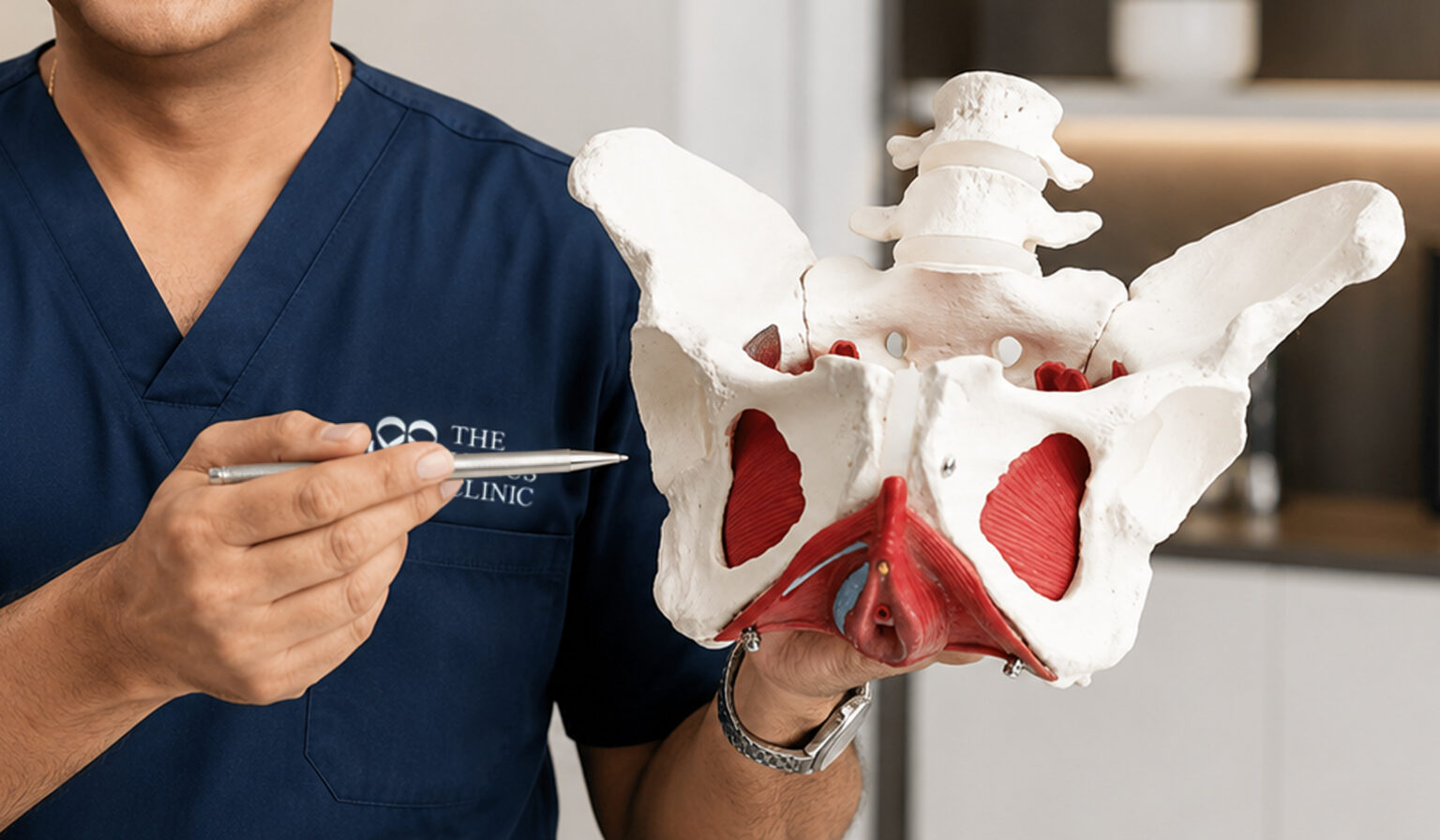
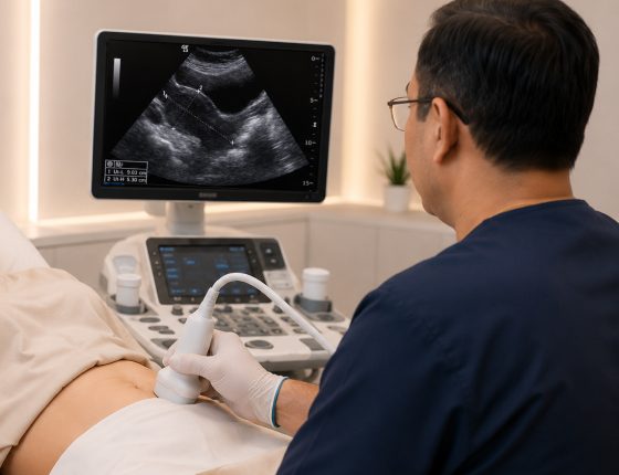
Diagnosing Pelvic Congestion Syndrome (PCS), now classified more broadly as Pelvic Venous Disorder (PeVD), requires a different type of investigation from a standard gynaecological ultrasound. Because PCS is a functional vascular condition — caused by abnormal blood flow in the pelvic veins, most commonly due to reflux through incompetent vein valves, though obstruction and other structural factors can also contribute — it can only be reliably identified using imaging that specifically assesses blood flow dynamics within the pelvic veins.

This page explains why standard scans often miss PCS, what a specialist diagnostic pathway involves, and what you can expect at each step. It is written to help you arrive at a consultation well-informed, with the right questions prepared.

Group 1993 Group 1993

It Is Not That Nothing Is Wrong — It Is That the Right Thing Has Not Been Checked

Frame 1975 Frame 1975

What a standard gynaecological ultrasound is typically not configured to do is evaluate the functional behaviour of pelvic veins.

Most routine protocols do not include vascular or Doppler assessment — meaning that even though ultrasound technology is capable of assessing blood flow direction and velocity, this evaluation is not part of a standard gynaecological scan.

Identifying PCS requires imaging that is specifically designed to measure the direction and velocity of blood movement within the pelvic veins.

Frame 25161 Frame 25161

There is a second reason standard scans often miss it.

When a patient lies flat — the standard position for most ultrasound, CT, and MRI examinations — the pelvic veins may partially decompress, and varicosities become less prominent.

The congestion that causes symptoms during the day, when a person is upright, and gravity is acting on the blood column, may look far less pronounced on a supine scan. Dilated veins can appear almost normal in this position. This is why some specialist protocols now incorporate upright or semi-erect positioning, or use specific breathing manoeuvres during ultrasound, to better replicate the conditions under which symptoms occur.

Frame 25161 Frame 25161

This is a systemic diagnostic challenge, not an individual clinical error.

Awareness of PCS as a distinct, assessable venous condition has grown significantly in recent years, but the specific imaging protocols required to detect it are not yet uniformly available or routinely requested in general clinical practice.

The Diagnostic Pathway for PCS

A Stepwise Assessment, Tailored to Each Patient

The diagnostic workup at The Venus Clinic follows a structured, stepwise approach. Not every patient requires every investigation. The pathway is guided by the findings at each stage and by the specific pattern of your symptoms.

The clinic offers four core assessments, described below in clinical sequence, from first-line to most detailed.

Step 1 — Trans-Abdominal and Transvaginal Duplex Ultrasound (The PCS Scan)

  • What it is: This specialist ultrasound is widely endorsed by pelvic venous specialists as the preferred first-line non-invasive investigation for PCS. It is meaningfully different from a standard pelvic ultrasound. Using Doppler technology, it assesses both the structure of the pelvic veins and, critically, the direction and velocity of blood flow within them. Where further confirmation is required, catheter venography remains the reference standard for definitive diagnosis.
  • The scan evaluates: the ovarian veins (left and right) for dilation and retrograde flow; the internal iliac veins and pelvic venous plexus; the left renal vein (for Nutcracker Syndrome); and the pelvic organs, to identify or exclude other conditions such as fibroids or ovarian cysts that may mimic or coexist with PCS.
  • Why it may differ from scans you have had before: The scan is conducted using a specific protocol developed for PCS assessment. It may include a Valsalva manoeuvre — you will be asked to bear down gently, increasing abdominal pressure — to provoke retrograde flow that may not be visible at rest. The scan may be performed with you partially upright or tilted, rather than lying completely flat.
  • What to expect: The scan involves two components: a trans-abdominal scan (the probe is placed on your abdomen, as with a routine ultrasound), followed by a transvaginal component (a small probe is gently placed internally). The transvaginal component allows clearer visualisation of the pelvic veins and organs. The procedure is typically performed in an outpatient setting. You will be asked to empty your bladder before the transvaginal portion. Discomfort, if any, is generally mild and brief.

Step 2 — Dynamic Pelvic Floor Ultrasound

  • What it is: This assessment evaluates the function of the pelvic floor muscles and identifies specific sites where venous blood may be leaking from the pelvis into the groin or legs. The pelvic floor and pelvic venous system are closely related anatomically and often affect one another — pelvic floor dysfunction and pelvic venous congestion frequently co-exist.
  • Why it matters: Understanding both allows the care team to plan management that addresses all contributing factors, not only the venous anatomy. This assessment also informs the physiotherapy component of care.

Step 3 — Venous Incompetency Duplex Ultrasound

  • What it is: In some patients, pelvic venous reflux does not remain confined to the pelvis. Blood leaking backwards through pelvic veins can travel into the veins of the legs and groin, presenting as varicose veins — particularly in the inner thighs, back of the thighs, vulval area, or buttocks. These are sometimes called atypical varicose veins because they arise from a pelvic, rather than a leg, venous source.
  • What the scan does: The venous incompetency duplex maps the path of these abnormal veins from their pelvic origin into the lower limbs. This information is important for treatment planning, as leg varicose veins that arise from a pelvic source require a different approach from standard leg varicose vein treatment.

Step 4 — Tilt-Table Test (for Postural Orthostatic Tachycardia Syndrome / POTS)

  • What it is: Postural Orthostatic Tachycardia Syndrome (POTS) is a condition of the autonomic nervous system in which blood pressure and heart rate do not regulate normally when moving from lying to standing. It produces symptoms including light-headedness, rapid heartbeat, brain fog, easy fatigue, abdominal bloating, and leg swelling.
  • Why it is relevant to PCS: Many women with PCS are also found to have features of POTS, and vice versa. Emerging evidence suggests that abnormal venous pooling may contribute to the circulatory instability seen in POTS when upright, though the precise relationship between the two conditions remains an active area of research. Identifying POTS alongside PCS is clinically important because it changes the scope of management.
  • What the test involves: You will lie on a motorised table that is then tilted to bring you to a near-upright position. Your heart rate and blood pressure are monitored throughout. The test identifies whether symptoms of autonomic instability are triggered by the postural change.

Step 5 — Further Imaging: CT Venography, MR Venography, and Catheter Venography

When further imaging is considered, for some patients, the duplex ultrasound assessment provides sufficient information to proceed with management planning. For others — particularly where anatomy is complex, where a compression syndrome such as Nutcracker Syndrome or May-Thurner Syndrome is suspected, or where a procedural intervention is being planned — additional imaging is indicated.

  • CT Venography: CT venography uses computed tomography with an injected contrast agent to produce detailed cross-sectional images of the pelvic venous anatomy. It is particularly useful for mapping the extent of dilated veins, identifying anatomical compression variants, and assessing collateral vessels. It involves exposure to ionising radiation and an iodine-based contrast agent. Kidney function is typically checked before the scan, as the contrast agent is cleared through the kidneys.
  • MR Venography: Magnetic resonance venography achieves similar anatomical mapping without ionising radiation. It is particularly useful for assessing soft tissue structures alongside the vascular anatomy and for evaluating pelvic organs in detail. It requires the injection of a gadolinium-based contrast agent. Patients with significantly impaired kidney function require specific consideration before gadolinium is used.
  • Catheter Venography — The Reference Standard: Catheter venography is the established reference standard for definitive assessment of pelvic venous reflux. A thin, flexible catheter is guided through a small puncture site — typically in the neck or groin — into the pelvic veins under real-time X-ray guidance. Contrast dye is injected directly into the veins, allowing the specialist to observe blood flow patterns directly, measure vein diameters against established diagnostic thresholds, and confirm whether venous reflux meets the criteria for PCS.

The procedure is performed under local anaesthesia with light sedation. Most patients describe a feeling of pressure rather than pain. A small bruise or tenderness at the puncture site is common and typically resolves within a few days.

In some cases, if the diagnosis is confirmed during catheter venography, the specialist may proceed to embolisation treatment in the same session. This decision is always discussed with the patient in advance. The diagnostic and treatment steps are distinct, even when they occur during the same procedure. Where this is a possibility, it will be explained clearly before you consent.

Preparing For Your Appointment

Frame 2147223881 Frame 2147223881

What to Bring

Arriving prepared makes the initial consultation more efficient and ensures your specialist has the full clinical picture from the outset. Bring the following, where possible:

  • A written summary of your symptoms: when they started, what they feel like, where they are located, and what makes them better or worse. Pay specific attention to whether symptoms change with posture (worse when standing, better when lying down) or with your menstrual cycle.
  • Reports and images from all previous investigations — including scans reported as normal. Even normal results provide useful context, as normal imaging findings do not exclude PCS.
  • A list of all current medications and supplements, including anticoagulants, antiplatelet agents (such as aspirin), NSAIDs, and herbal or nutritional supplements, as some of these may need to be reviewed or paused before catheter-based procedures.
  • Details of any previous treatments tried, including medications, hormonal therapies, physiotherapy, or surgery.
Frame 2147223882 Frame 2147223882

Why You May Be Asked to Repeat Some Investigations

Previous scans were likely performed with you lying flat, without Doppler assessment of vein blood flow dynamics, and without provocation manoeuvres.

If your specialist determines that a PCS-specific duplex ultrasound with Doppler assessment and positional provocation is needed, this is not necessarily a duplication of what you have already had — it is a different investigation designed to assess what the earlier scans were not designed to show. In some cases, prior imaging may still provide useful context and be reviewed alongside any new assessment.

Specialist Conversation Checklist

Knowing what to ask helps you make the most of your appointment and ensures you leave with a clear understanding of the next steps.

About your symptoms:


  • Based on what I have described, does my symptom pattern suggest a venous cause is worth investigating?

  • What aspects of my history make you more or less likely to suspect PCS?

  • Are there other conditions you would want to rule out first, and how would you do that?

About the diagnostic process:


  • Which investigations do you recommend for me, and in what order?

  • Will the duplex ultrasound include a Doppler assessment of blood flow direction?

  • Will I need to be in an upright or tilted position during the scan?

  • What diagnostic criteria are used to confirm PCS — what specifically would you need to see?
  • How will you differentiate PCS from endometriosis, fibroids, or another pelvic condition?

About results:


  • How will my results be communicated — in a follow-up appointment, by phone or email?

  • If my scan appears normal, what would the next step be?

  • Under what circumstances would you recommend catheter venography?

About requesting a referral (if seeing a general practitioner or gynaecologist first):


  • Could you refer me for a trans-abdominal and transvaginal duplex ultrasound specifically assessing for pelvic venous reflux?

  • Is a referral to a vascular specialist appropriate, given my symptom history?

Note: You do not need a GP referral to make an initial enquiry at The Venus Clinic. You may contact the team directly to discuss your symptoms and ask

Safety And What To Expect After Each Investigation

4.9 stars (500+ Google Reviews)
Frame 25157

Duplex Ultrasound (trans-abdominal and transvaginal)

No radiation, no contrast agent, no needles. Safe to repeat. No recovery time needed — you may return to normal activities immediately.

Frame 2147223884

Dynamic Pelvic Floor Ultrasound

Same safety profile as duplex ultrasound. Non-invasive and well-tolerated.

Frame 2147223883

Venous Incompetency Duplex

Same safety profile as duplex ultrasound. No special preparation or recovery needed.

Frame 2147223886

Tilt-Table Test

No needles or radiation. Some patients feel light-headed, dizzy, or nauseated during the test — this is part of how the autonomic response is assessed. In some cases, a brief loss of consciousness may occur; clinical staff are present throughout and will immediately return the table to a flat position if needed. Your medical history will be reviewed beforehand to confirm that the test is appropriate for you. You should arrange to be accompanied if you feel unsteady afterwards.

Frame 25156

CT Venography

Involves ionising radiation and an iodine-based contrast agent. Kidney function is checked beforehand. Mild reactions to contrast are possible; severe reactions are uncommon. A brief rest period after the scan is recommended.

Frame 2147223886-2

MR Venography

No radiation. A gadolinium-based contrast agent is used; special consideration is required for patients with impaired kidney function. Patients who experience claustrophobia should inform the team in advance. No recovery time is needed, but you should not drive if sedation has been offered.

Frame 2147223886-1

Catheter Venography

Minimally invasive. Performed under local anaesthesia with light sedation. You will be awake throughout. A bruise or mild tenderness at the puncture site is common. Rest is advised for the remainder of the day. Your specialist team will provide written post-procedure instructions, including specific signs to watch for — such as increasing pain, swelling, or fever — that would warrant contacting the clinic.

Understanding Your Results

How a Diagnosis Is Made

A diagnosis of PCS is not made from a single measurement. It involves the careful correlation of multiple clinical findings with your reported symptoms. Key elements your specialist will interpret include:

Ovarian vein diameter Veins dilated beyond published diagnostic thresholds — which vary by modality but typically include ovarian vein diameter above 6–8 mm and parauterine veins above 4 mm — may indicate abnormal venous pressure. Diameter alone is not sufficient to make a diagnosis; it must be interpreted alongside flow velocity and reflux findings.
Reflux pattern and duration The direction, duration, and velocity of retrograde blood flow through incompetent valves during provocation testing. A reflux duration of more than one second during Valsalva, combined with low flow velocity, is considered diagnostically significant.
Distribution of pelvic varices The location and extent of dilated veins within the pelvis, and whether they extend into the legs or vulval area.
Pelvic floor function Whether pelvic floor dysfunction is present alongside venous findings, as the two conditions frequently co-exist and may influence the overall management plan.
Symptom correlation Whether the imaging findings account for the specific symptom pattern you have described.

Although enlarged pelvic veins and pain are the hallmark features of PCS, asymptomatic women have also been found to have pelvic varicosities, which means that imaging findings must always be interpreted in the context of your symptoms. It is the functional assessment — the presence of retrograde flow correlated with symptoms — that forms the basis of diagnosis.

If initial investigations are inconclusive, your specialist may recommend repeat testing under different conditions, additional imaging, or further evaluation to exclude concurrent contributing conditions.

Frame 1976 Frame 1976

The Next Step:
From Diagnosis to Management

Once the diagnostic workup is complete, the focus shifts to planning an appropriate management approach.

This may range from conservative measures and physiotherapy to minimally invasive vascular procedures, depending on the findings and the severity of your symptoms.

Frequently Asked Questions

Standard pelvic ultrasounds and gynaecological MRI scans are designed to assess organ structure, not venous blood flow dynamics. PCS-specific imaging requires Doppler assessment of the direction and velocity of blood flow within the pelvic veins, combined with provocation techniques such as a Valsalva manoeuvre, and may need to be conducted with the patient in an upright or tilted position rather than lying flat. This is a different investigation from a routine scan. A result of “normal” on a standard pelvic scan does not exclude PCS.

A referral is not required to make an initial enquiry. You may contact the team directly by phone, WhatsApp, or via the enquiry form on the Contact page.

This varies by individual. The PCS duplex ultrasound can often be arranged within a short period after an initial consultation. Further investigations, if needed, depend on scheduling and on the findings at each stage. Some patients complete their workup within a few weeks; others may require a longer sequence of assessments.

Not all patients require catheter venography. Many can be assessed fully with the duplex ultrasound and, where needed, cross-sectional imaging. Catheter venography is recommended when the findings from earlier investigations support its use or when a procedure is being planned in the same session.

Yes. The symptom overlap between PCS, endometriosis, and uterine fibroids is significant, and the conditions frequently co-exist. This is clinically important: in some women, an existing diagnosis of endometriosis or fibroids may not fully account for all their symptoms, and a concurrent venous component may be missed without targeted venous imaging. Identifying all contributing factors allows for a more complete and effective management plan.

The procedure is performed under local anaesthesia with light sedation. Most patients describe a sense of pressure rather than sharp pain during the procedure. Mild soreness at the catheter insertion site is common in the days following and typically resolves within a week.

A confirmed diagnosis, where venous reflux and associated symptoms are identified, provides the clinical basis for a structured management plan. This may involve targeted pelvic physiotherapy, conservative medical management, or minimally invasive vascular treatment, tailored to your individual findings. Individual responses to treatment vary and are discussed in detail with your specialist.

Costs vary depending on which investigations are required for your specific clinical picture. An accurate estimate can only be provided after an initial consultation. The Venus Clinic team can discuss this with you at your appointment. We recommend checking with your insurer regarding coverage prior to your consultation, as individual insurance policies vary.

Duplex ultrasound, dynamic pelvic floor ultrasound, and MR venography do not involve ionising radiation. CT venography and catheter venography under fluoroscopic guidance do involve ionising radiation. As with all imaging using radiation, doses are managed according to the ALARA (As Low As Reasonably Achievable) principle — the minimum dose necessary to obtain diagnostically adequate images.

Not Sure Where to Start?

If you have been experiencing persistent pelvic pain and investigations have not provided a clear explanation, a specialist assessment designed specifically to evaluate pelvic venous function may offer clinical information that has not yet been explored.

image 12520 mobile doc

Led by Dr Sriram Narayanan

Senior Vascular and Endovascular Surgeon

The Venus Clinic is led by Dr Sriram Narayanan (Dr Ram), a Senior Vascular and Endovascular Surgeon with over two decades of clinical experience in vascular surgery in the United Kingdom and Singapore.

  • Bachelor of Medicine and Master of Surgery (Bombay)
  • Fellow of the Royal College of Surgeons (General Surgery, Glasgow)
  • Dip Laparoscopic Surgery (France)

Dr Ram was among the first surgeons in Singapore to develop vein stenting, intravascular ultrasound (IVUS), and duplex ultrasound assessment protocols for Pelvic Congestion Syndrome. He has published extensively on vascular disease and has served as a faculty member and trainer at international vascular and vein conferences, including training programmes in the UK and Asia focused on pelvic duplex ultrasound for PCS.

Learn more about Dr Ram

Speak With Our Team

If you would like to discuss your symptoms or enquire about a consultation, please contact us by phone, WhatsApp, or email. You may also complete the enquiry form on our Contact page. A GP referral is not required to make an initial enquiry.

    VISIT US

    Our Clinic

    Our Clinic – Dr Sriram Narayanan

    image 12520

    Led by Dr Sriram Narayanan

    Director , Senior Consultant Vascular & Endovascular Surgeon

    Mount Alvernia Hospital, #01-03 Medical Centre A,
    820 Thomson Road, Singapore 574623

    +65 6219 3108 (Tel)
    +65 8666 9639 (Whatsapp)

    Mon – Fri: 8:30 AM – 5:30 PM
    Weekends & Public Holidays: Closed

    +65 8666 9639